What is Iridotomy?
Laser iridotonomy (Nd: YAG laser, Argon laser) provides intraocular fluid passage to the anterior part of the eye by opening the iris, thus creating a pressure balance in the anterior and posterior parts of the eye, decreasing the increased intraocular pressure to normal level, reducing severe pain and recurrent It is the process of preventing attacks.
In people who have undergone cataract surgery, the membrane behind the lens placed inside the eye becomes dull over time. This dullness slowly decreases vision. The patient feels as if cataract is starting again. After enough time from the operation, YAG laser application (capsulotomy) must remove this dullness. The application is actually a light therapy and it is possible to apply it without contacting the eye with any device or instrument. It is a process that takes seconds and is completely painless. The application is performed while the patient is sitting on the examination chair.
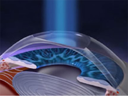
In the treatment of some types of glaucoma (eye pressure), eye pressure is reduced by performing iridotomy with YAG laser. This process is the creation of a millimetric opening in the iris tissue that gives the eye color.
What is Photocoagulation?
Argon laser is a type of ion laser that is filled with argon gas and generates light at blue / green wavelengths. In cases of refraction, the laser is welded to abnormal vessels. Because the patient adapts himself to daily life more quickly.
What is Vitreous Hemorrhage?
The majority of the eyeball is filled with a liquid in the consistency of the egg flow called the vitreous. The bleeding that occurs into this liquid is called intraocular bleeding (vitreous hemorrhage). Although there are many reasons for intraocular bleeding, most of them are vascular reasons. Intraocular bleeding is common, especially in the advanced stages of diabetic retinopathy due to diabetes. In addition, it may develop after the obstruction in the retinal vessels. It may occur with systemic diseases such as leukemia due to indirect or direct trauma as well as the presence of retinal tears as a result of the failure of retinal vessels due to premature birth.The most common causes of intraocular bleeding are listed below:
Diabetic retinopathy (32-54%), With retinal tear (12-44%),Posterior vitreous detachment (412%), Retinal detachment (7-10%)şSickle cell anemia (1-6%), Retinal macroaneurysm (1-7%)
Mourning yellow spot disease (1-5%), With intracranial hemorrhages (1%),Trauma (12-19%), Retinal vein occlusion (4-16%)
If intraocular bleeding remains in the eye for a long time, regardless of the reason, it may cause some complications beyond the damage caused by the underlying disease. One of them is the release of iron element in blood cells and their absorption by the cells in the retina and other layers, resulting in some toxic effects. These toxic effects prevent the healthy functioning of retinal cells. Another effect is glaucoma, which increases the eye tension by obstructing the outer pathways of the eye by the damaged blood cells circulating in the vitreous.
WHAT IS THE TREATMENT OF INTRAOCULAR BLEEDING?
The treatment of bleeding depends on the underlying cause. If the cause is retinal tear or retinal detachment, urgent surgery and / or laser treatment is required. If it has occurred due to a systemic vascular disease such as diabetes, it should be followed up with frequent checks every 1-2 weeks by applying head-up / vertical rest, and if there is no opening in a period of 1-2 months, a bleeding removal surgery called vitrectomy should be applied.
In the meantime, the patient should be consulted in terms of systemic diseases such as diabetes, hypertension and blood diseases, and blood values in this regard should be normalized.
In cases where the visual center called macula is healthy, the visual gain after surgery is also very pleasing. However, in diseases affecting the macula, such as diabetic retinopathy and age-related macular degeneration, vision expectancy is relatively limited.
What is Retinal Detachment?
It develops due to tears or holes in the retina. It is frequently seen in patients with high myopia. It can occur at any age, mostly middle age and above.
The retinal layer is stretched as the anteroposterior diameter of the eye increases and the stretch area on it begins to thin and deteriorate. In some familial or degenerative diseases and infections, thinning and deterioration may occur around the retina. Meanwhile, for the same reasons, the vitreous gel also begins to lose its homogeneity and deteriorate, the gel consistency changes and slowly separates from the retina. This separation is called vitreous detachment. In the meantime, as the vitreous tissue, which shrinks and becomes opaque in places, passes through the visual axis in the eye, it is perceived by the person as flying flies or smoke screens. If retinal detachment is not treated immediately by an experienced physician in the retinal area, it may cause partial or complete vision loss.
Retinal Detachment Causes and Symptoms
Retinal diseases can result in permanent blindness if left untreated. Symptoms such as light flashing, flying fly, sudden vision loss can be the harbinger of an important eye disease such as retinal detachment. In its treatment, it is possible to prevent results that may lead to vision loss with early diagnosis, detailed examination, timely and most importantly correct treatment. Retinal surgeries are important operations that require the use of high technology with great sterilization measures, otherwise the result may result in vision loss.
Central vision is lost when the macula (the visual center of the eye) detaches from the underlying tissue. In long-term detachments, intraocular balances are disturbed and the eyeball begins to shrink. Sudden, severe or penetrating blows to the eyes can cause detachment. In diabetes and some degenerative diseases, detachments due to traction may develop by forming bands that pull the retina in the vitreous. Although rarely, detachment can develop without any tears in the eye, in some infections, tumors or blood pressure crises that occur especially during pregnancy.